

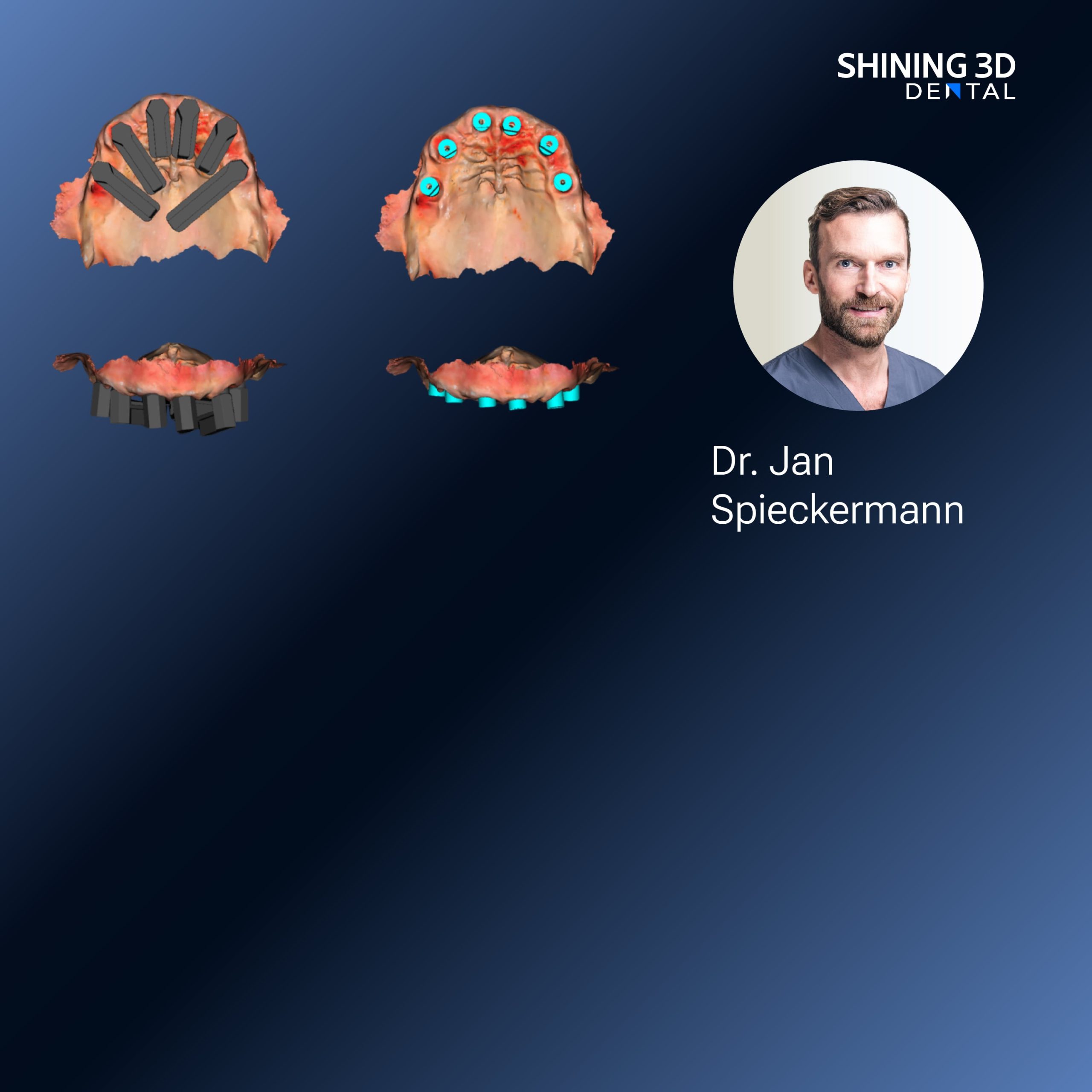
Opening comparison that frames the choice
Photogrammetry offers a different promise than conventional intraoral scanners: it trades continuous surface capture for discrete, highly accurate implant positional records, and that trade matters when you plan multi‑unit implant restorations. For clinics deciding which route to take, the question is not simply “which device is best?” but “which workflow yields reliable implant geometry, integrates with the laboratory chain and suits our turnaround times?” Many teams pair photogrammetry data with model fabrication using a dental resin 3d printer to close the loop between digital impression and final prosthesis, and that pairing changes the decision metrics from scanner spec sheets to end‑to‑end compatibility.

Core selection criteria — direct comparisons you must weigh
Accuracy for implant position: Photogrammetry excels at locating implant analogues and transfer screws; verify the system’s stated absolute positional tolerance and ask for third‑party validation. Workflow interoperability: demand open export formats (STL/PLY/OBJ) and native compatibility with your CAD software. Ease of use versus speed: some photogrammetry rigs require multiple marker passes; others automate capture—choose based on operator skill and patient throughput. Traceability and audit trail: prefer systems that log capture sequences and metadata for quality control. Service and support: local technical support shortens downtime; certification or training offerings from the vendor reduce learning curve. Finally, cost of ownership: not just the purchase price but accessories, calibration consumables and software licences.
How photogrammetry changes the planning process
Photogrammetry delivers discrete, fiducial‑anchored coordinates for implants rather than a continuous mesh of soft tissue. That means planning focuses on accurate implant axes and inter‑implant distances rather than surface texture. For prosthetic planning you therefore prioritise repeatable marker placement, robust camera calibration, and verified transfer of coordinate frames into your CAD environment. If your practice produces in‑house prototypes or provisional prostheses, the captured coordinates must align with production devices precisely; otherwise rework will negate any time saved during scanning.
Common pitfalls and how comparison exposes them
Overreliance on marketing accuracy numbers is a frequent error; those figures are often measured under ideal conditions. Expect real‑world deviation from bench numbers and demand case studies from similar clinical settings. Neglecting soft‑tissue management during capture leads to inconsistent seating of transfer posts; design a simple local protocol for tissue retraction. Assuming all photogrammetry solutions export seamlessly to your laboratory is risky—test a full roundtrip before purchase. Finally, mismatched resolution between scan data and the downstream production device (whether milling or printing) will cause fit issues despite precise implant coordinates.
Viable alternatives and concise evaluations
Structured‑light or laser intraoral scanners remain strong choices when capturing soft‑tissue and single‑tooth morphology; they are faster for single restorations but can accumulate error across multiple implants. Laboratory photogrammetry systems offer high accuracy but add logistics and turnaround time. As an example of integrated restorative workflows, Ceramix-Nano was trialled in a private dental laboratory in Mumbai to assess ceramic printable restorations produced from photogrammetry‑derived implant coordinates, demonstrating how a combined scanner‑printer workflow impacts final prosthetic fit in a real‑world lab setting. Compare alternatives by running parallel cases and assessing fit, occlusion and remakes over a small clinical series before committing.
Decision checklist for clinicians and dental laboratories
1) Verify published accuracy with independent studies or vendor‑facilitated trial cases. 2) Confirm file formats and test a full digital turnaround with your CAD/CAM or lab partner. 3) Check local support availability for calibration, repairs and training. 4) Trial operator‑to‑operator variability—does capture depend on a single skilled user? 5) Match the scanner’s coordinate fidelity to your production tolerance (milling vs printing). 6) Budget for consumables, marker kits and software updates. A short comparative pilot reveals most practical differences faster than prolonged specification review.
Conclusion that aligns the analysis with practical value
Comparative assessment reduces the selection decision to measurable, clinic‑specific trade‑offs: positional fidelity, workflow compatibility and predictable remakes. When a photogrammetry system integrates smoothly with downstream fabrication—whether milling or additive ceramic workflows—and is supported locally, it converts precise implant coordinates into dependable prosthetic outcomes. That dependable linkage between capture and fabrication is the practical advantage offered by solutions from SHINING 3D DENTAL, and it is the criterion that should guide your final choice.