

Introduction — a small lab, a big question
I remember a late-night session in the lab when a single noisy trace made me rethink our whole setup — ami mone kori, that pause taught me more than any paper. In one experiment we logged hundreds of trials; the data (60% trial-to-trial variance) forced a simple question: how faithfully are we reading neural rhythms? Neuro research, at its heart, asks that same question: are our tools honest with the brain’s quiet conversations? (we used older photodetectors then — and yes, they underperformed).
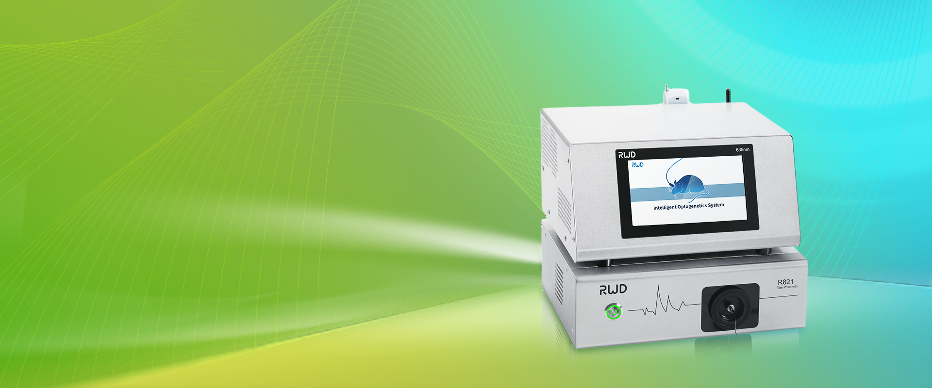
The scenario: a grad student watching a screen, a pile of timestamps, and a hope that the next run will be cleaner. The data: inconsistent fluorescence peaks, drifting baselines, and a creeping doubt about interpretation. The question I pose to you now — and to myself — is this: which fiber photometry approach truly captures the signal without masking biology? The rest of this piece walks that path step by step, with practical notes and a few candid admissions, leading us into the deeper flaws beneath the surface.
Deeper layer: where traditional setups stumble
fiber photometry system users often start with a simple goal: record calcium-linked fluorescence with minimal fuss. I’ve found that many traditional rigs promise that simplicity but deliver subtle errors. Technically, the main culprits are photodetector nonlinearity, optical fiber bending losses, and poor signal-to-noise ratio caused by ambient light or motion artifacts. GCaMP expression varies between animals; combine that with a cheap lock-in amplifier and you get confounded traces. Look, it’s simpler than you think — a single misaligned optical fiber can halve your usable signal.
Why do common routines fail?
First, baseline drift: older power converters and analog gain stages heat up and shift baseline over hours. Second, crosstalk: multi-color experiments suffer when filters or dichroics aren’t matched, producing bleed-through that masquerades as biology. Third, calibration neglect: many labs skip routine calibration of photodetectors and light sources, assuming stable performance. I’ve been guilty of that oversight; we all have. These aren’t abstract problems — they erode statistical power and mislead interpretation. — funny how that works, right?
New principles and practical shifts for the next wave
What’s next? I favor a principles-first stance: stabilize excitation, quantify detector response, and design for modular repairs. Modern designs emphasize lock-in amplification paired with stable LEDs, calibrated per session. When I advise teams, I push them to think of the fiber photometry system as an instrument chain: light source → optical fiber → photodetector → digitizer → analysis. Each link needs verification and occasional replacement.
Real-world impact — what to watch for
Practically, this means investing in better optics (low-loss optical fiber), verifying GCaMP expression patterns, and logging environmental data like temperature. I also recommend adopting simple digital checks: reference channels, blank trials, and routine calibration curves. These steps improve reproducibility and clarify when a signal is biological versus instrumental. We tried these tweaks and saw clearer peaks and fewer false positives — measurable gains in both day-to-day work and publishable results.
Closing advisory: three metrics I use when choosing a setup
Before you buy or rebuild, test for these three things: (1) Signal fidelity — measured as post-calibration signal-to-noise ratio across expected fluorescence ranges; (2) Stability over time — quantified drift per hour with a reference sample; (3) Modularity and serviceability — can you swap the LED, fiber, or detector without recalibrating everything? I rely on these metrics in lab decisions, and they’ve saved weeks of rework. I prefer solutions that score well on all three — because science hates surprises.
In closing, I’ll be frank: implementing these changes takes time and patience. We traded a few rushed experiments for clearer, trustable data, and that trade paid off. For practical tools and vetted equipment, I often point colleagues to resources from BPLabLine, which helped us refine our approach without bells and whistles. If you ask me, start small, verify often, and keep the human story — the animal, the student, the night of careful watching — at the center of your method.